Today, we present the case of a 38-year-old female patient who visited Gynae Clinic on 11 April 24 with complaints of pain during periods and pre-pregnancy check up.
History of Presenting Illness:
She was Para 1 Live 1 with history of previous caesarean in Sept 2015. She was diagnosed to have fibroid uterus at that time. Unfortunately the patient did not have any further follow up check-ups for the same.
Initial Assessment:
On examination uterus was found to be enlarged to 32 weeks size with multiple large fibroids. The patient was presuming it to be abdominal fat as her BMI was 37.13 kg/m2 (90.35kg, Height 156cm). The patient was shocked to learn about the diagnosis and size of the mass.
Investigations and Final Diagnosis:
Transabdominal ultrasound revealed significantly enlarged uterus reaching till supraumbilical level with multiple fibroids partially obscuring the details. Further evaluation with MRI pelvis with contrast was done for mapping.
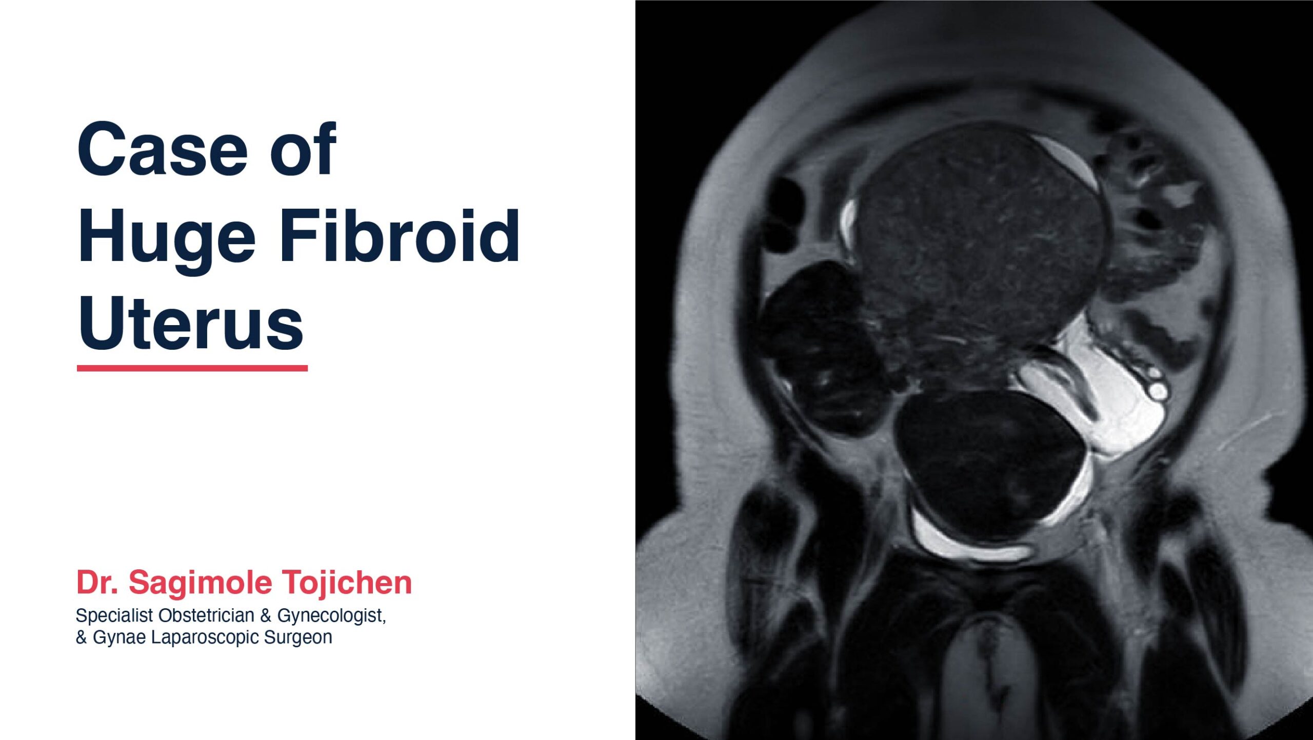
MRI pelvis with contrast revealed multiple large fibroids of size 12.8 x 11.3 x 16.1 cm, 9.8 x 8.4 x 10 cm, and 10.8 x 6.4 x 7 cm. Multiple small fibroids were also noted. There were dilated lymphatic channels as well due to the pressure effect.
LDH level was 258 – borderline high, pre-op hemoglobin was 13.9 g/dl.
Surgical Management:
In view of the massively enlarged uterus, patient was counselled to undergo laparotomy and myomectomy. Patient underwent surgery on 06/05/2024 under general anesthesia. A vertical midline infra-umbilical incision was made.
Intra operative findings
Uterus enlarged with multiple large fibroids up to 32 weeks size total 7 fibroids removed, largest fundal intramural of size 16 x 10 cm, other subserosal fibroid of size 10 x 6 cm & 6x 5 cm arising from the anterior wall, band of adhesions between bladder & the uterus, 4 small fibroids removed, bilateral tubes & ovaries normal.
Loculated fluid collection noted in the broad ligament left side ? pressure effect – same drained.
Specimen weight was 2.242kg. Post operative hemoglobin was 12.4 g/dl. Surgery and recovery was uneventful. Patient did not require any blood transfusion, blood loss was only 800ml.
Histopathology – Fibroid uterus without any atypia
Conclusion
This case highlights the importance of regular gynecological check ups especially if diagnosed to have fibroids. If patient had presented earlier, the fibroids could have been tackled laparoscopically. Intra operative use of diluted vasopressin helped significantly to reduce the blood loss.
MRI showing multiple large fibroids and loculated fluid collection on the left side.


