Introduction:
A 15-year-old female presented to the Ophthalmology clinic approximately 3 months ago with a history of outward deviation of the left eye along with double vision.
History of Presenting Illness:
Initially, the outward deviation of the left eye was intermittent, noticed only when she was tired, inattentive, sick, or exposed to bright sunlight. Over time, the left eye deviation progressed to being present for more than 50 percent of the waking hours, accompanied by worsening double vision or diplopia. This indicated that the Exotropia was becoming decompensated.
Initial Assessment and Differential Diagnosis:
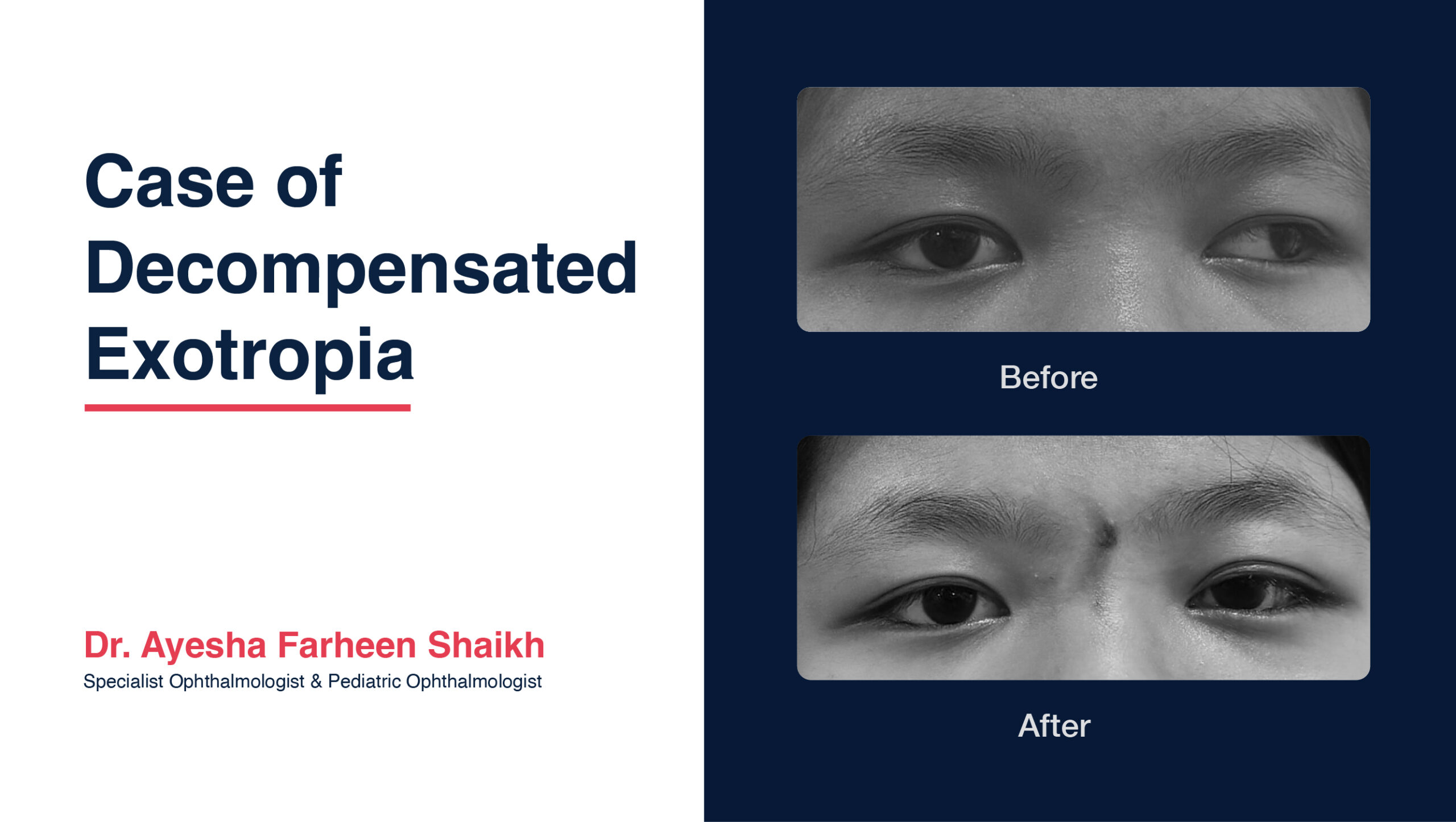
A complete ocular examination was performed, including anterior and posterior segment examinations, as well as motor and sensory evaluations for Exotropia. The best-corrected visual acuity for both eyes was 6/6 and N6. The corneal reflex showed 45-degree Exotropia in the left eye. The cover test demonstrated that the Exotropia was alternating with right eye dominance and that control of the Exotropia was fair. Ocular movements were full in all cardinal gazes. The prism cover test revealed approximately 85 prism diopters of left eye Exotropia with tenacious proximal fusion. The sensory examination using Worth four-dot testing indicated diplopia, as the patient saw 5 circles. Both anterior and posterior segments of the eyes were normal. Systemic and neurological examinations were normal, ruling out any neurological causes for the left eye Exotropia.
Investigations and Final Diagnosis:
After thorough evaluation, the decision was made to surgically correct the squint. Major concerns included the active tenacious proximal fusion, decompensation of the deviation, and the development of diplopia. Considering these factors and the parents’ concerns, the surgery was planned for the affected eye only, allowing room for a possible secondary procedure on the better eye in the future if needed. The child underwent a left eye lateral rectus recession (9.5 mm) and medial rectus resection (6.5 mm) under general anesthesia. The surgery was uneventful, and the patient tolerated the procedure well.
Post-Operative Examination:
Post-operatively, the patient was examined the next day. The corneal reflex showed approximately 5-degree Exotropia in the left eye, the sutures appeared intact, and the patient no longer complained of double vision. The patient was discharged with instructions for post-operative care.
On the 10th post-operative day, the patient was examined in the OPD. The corneal reflex was now central, and the patient had achieved optimal single binocular vision. Both the patient and her parents were extremely happy and satisfied with the surgical outcome. She has been advised to continue her post-operative care and attend regular follow-ups.
Conclusion:
This case highlights the importance of regular eye check-ups. Not all squints require surgical correction; such decisions should be made after thorough examination. For those requiring surgical intervention, time and expertise are crucial, as they impact both the surgical motor outcome in terms of optimal ocular alignment and the sensory outcome in terms of achieving single binocular vision.